
Clinical Workflow Transformation

Cardiac SPECT/CTClinical Workflow Transformation

I advanced NM Myocardial Perfusion imaging through soft-tissue attenuation correction and an optimized clinical workflow.

JHAHNuclear MedicineClinical Systems

01Before

Conventional Cardiac SPECT

Conventional myocardial perfusion imaging could require additional views to address breast and diaphragmatic attenuation artifacts.

Extra Images → More Examination Time

More time for the patient on the imaging table.
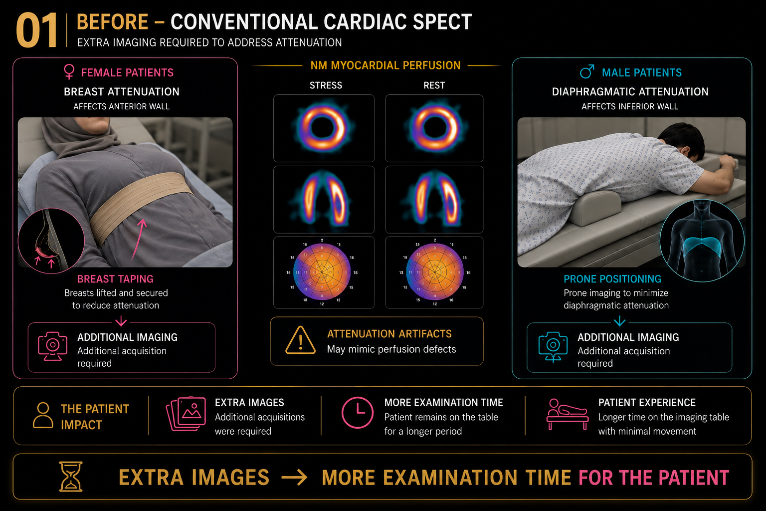
Conventional Cardiac SPECT workflow requiring additional imaging for attenuation artifacts View full size
Before — Conventional Cardiac SPECT

02The Transformation

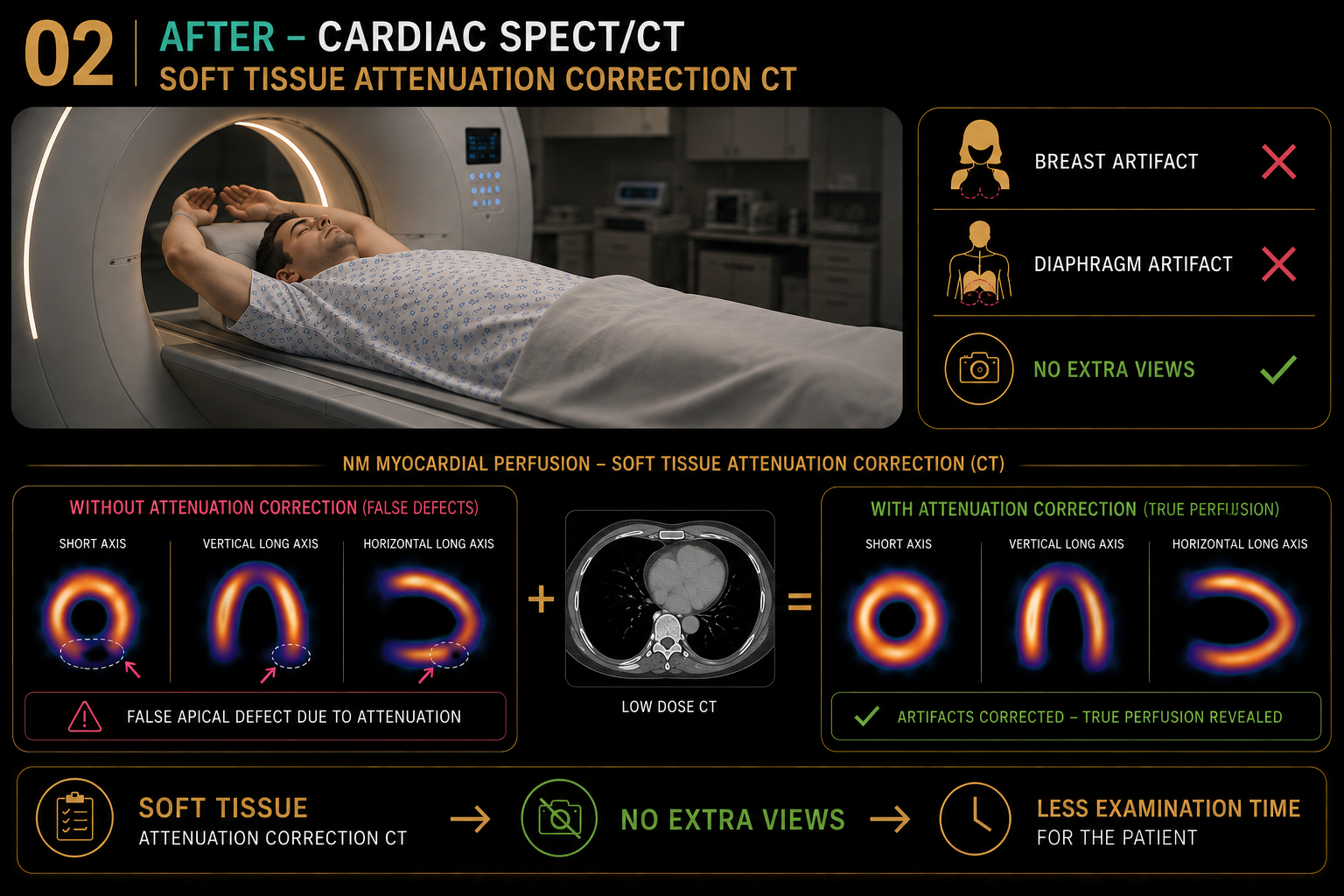
Introducing Cardiac SPECT/CT

I transitioned cardiac imaging to SPECT/CT and integrated CT-based soft-tissue attenuation correction into the myocardial perfusion workflow.

Soft-Tissue Attenuation Correction

Breast artifact ✕ • Diaphragm artifact ✕ • No extra views

Cardiac SPECT CT workflow with soft-tissue attenuation correction View full size
After — Cardiac SPECT/CT attenuation correction

The Result

Fused NM + CT Imaging. Computed Attenuation Correction. More Accurate Results. Less Imaging Time.

My implementation combined fused NM and CT imaging with computed attenuation correction, improving diagnostic accuracy while reducing imaging time for the patient.